The Gram Stain Provides a Lot of Clinically
Nearly all clinically important bacteria can be detectedvisualized using Gram staining method the only exceptions being those organisms. This is based on cell.

Gram Negative Microorganism Flow Chart Microbiology Microbiology Study Medical Laboratory Science
The name comes from the Danish bacteriologist Hans Christian Gram who developed the technique in 1884.

. Here are our formulas for Gram stain reagents. There are two main categories of bacterial infections. Gram stains may also be used to check for bacteria in certain body fluids such as blood or urine.
That exists almost exclusively within host cells ie. The Gram stain provides a lot of clinically useful information but it wont give you all the information you need for identification and treatment. About which of these do you NOT learn anything from the Gram stain.
Both gram-positive and gram-negative cells have. Gram-positive bacteria and Gram-negative bacteria. Catalase test tests for the reaction of hydrogen peroxide into oxygen and water indicating the presence of catalase indole test determines production of indole from tryptophan Which stains are used to visualize structures in the membrane Acid-fast and Gram The Gram stain stains peptidoglycan and the acid-fast stain stains mycelia acid which are both in the cell wall what.
Intracellular bacteria eg Chlamydia. A set of Good Clinical Laboratory Practice GCLP standards that embraces both the research and clinical aspects of GLP were developed utilizing a variety of collected regulatory and guidance material. Gram Staining appearance differs.
The gram stain could be done in 30 minutes stat or typically within 2-4 hours. Gram staining differentiates bacteria by the chemical and physical properties of their cell. About which of these do you NOT learn anything from the Gram stain.
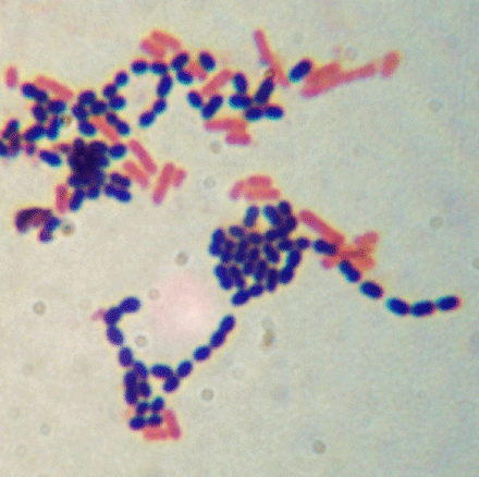
A Gram stain is a test that checks for bacteria at the site of a suspected infection such as the throat lungs genitals or in skin wounds. The Gram stain involves staining bacteria fixing the color with a mordant decolorizing the cells and applying a counterstain. The gram stain is used as the basis for treatment and is potentially life saving in critically ill patients with meningitis or sepsis.
A unique BD manufacturing process reduces the presence of artifactsa chemical precipitate sometimes mistaken for microorganisms in Gram stain preparations. Details vary from one Gram stain protocol to another mainly in the timing and the composition of the decolorizing agent. A clinician cannot wait for the complete work up of a C S 3-5 days to treat someone in septic shock.
Gloves laboratory safety glasses and a lab coat are recommended. Here are the advantages and disadvantages of Gram staining. The Gram stain provides a lot of clinically useful information but it wont give you all the information you need for identification and treatment.
This can help your doctor determine. The Gram stain provides a lot of clinically useful information but it wont give you all the information you need for identification and treatment. Gram stain or Gram staining also called Grams method is a method of staining used to classify bacterial species into two large groups.
About which of these do you NOT learn anything from the Gram stain. The categories are diagnosed based on. The Gram stain provides a lot of clinically useful information but it wont give you all the information you need for identification and treatment.
It gives quick results when examining infections. The genera Actinomyctes Arthobacter Corynebacterium Mycobacterium and Propionibacterium have cell walls particularly sensitive to breakage during cell division resulting in Gram-negative staining of these Gram-positive cells. The procedure is based on the reaction between peptidoglycan in the cell walls of some bacteria.
List of Advantages of Gram Staining. 1 See answer Roddyabeast8740 is waiting for your help. The Gram stain was originally devised by Christian Gram in 1884.
The end result is easier-to-read and accurate staining. The staining of these organisms result in an uneven or granular appearance DrTVRao MD Gram. In most cases Gram stains are performed on biopsy or bodily fluids when infection is suspected and they yield results much more quickly than other methods such as culturing.
The stan- dard Grams staining method can be used to differentiate intact morphologi- cally similar bacteria into two groups. The main benefit of a gram stain is that it helps your doctor learn if you have a bacterial infection and it determines what type of bacteria are causing it. Biology Thu Thủy 6 months 2021-07-21T0546450000 2021-07-21T0546450000 1 Answers 246 views 0.
We describe eleven core elements that constitute the GCLP standards with the objective of filling a gap for laboratory guidance based on IND. Add your answer and earn points. What is a Gram stain.
A Structure of the cell wall b Bacterial morphology c Susceptibility to antimicrobial drugs d The ability of the bacteria to process. The Gram stain provides a lot of clinically useful information but it wont give you all the information you need for identification and treatment. Within our state-of-the-art ISO 9000 manufacturing center is a dedicated production area for stain reagents.
The Gram stain provides a lot of clinically useful information but it wont give you all the information you need for identification and treatment. The ability of the bacteria to process nutrients which type of medium supports the growth of the widest range of bacteria. This stain is used to identify the presence of microorganisms in normally sterile body fluids cerebrospinal fluid synovial fluid pleural fluid peritoneal fluid.
About which of these do you NOT learn anything from the Gram stain. About which of these do you NOT learn anything from the Gram stain. The Gram stain provides a lot of clinically useful information but it wont give you all the information you need for identification and treatment.
The Gram stain is used to provide preliminary information concerning the type of organisms present directly from clinical specimens or from growth on culture plates. The primary stain crystal violet binds to peptidoglycan coloring cells purple. Gram stain reagents will stain skin and clothing.

2 4 Staining Microscopic Specimens Biology Libretexts Microbiology Microbiology Textbook Medical Terminology Study

Pdf Gram Stain And Ziehl Neelsen Stain Limited But Important Specific Diagnostic Tool

Gram Staining Procedure 20 Download Scientific Diagram
Gram Stain From A Male Urethral Swab Depiction Of Polymorphonuclear Download Scientific Diagram

Different Size Shape And Arrangement Of Bacterial Cells When Viewed Under Light Microscope Most Bacteria Appea Bacteria Shapes Microbiology Microbiology Lab

Fecal Gram Staining Of Phagocytosed Bacteria To Differentiate Methicillin Resistant Staphylococcus Aureus A Case Report Journal Of Infection And Chemotherapy

Management Of Acute Pancreatitis American College Of Gastroenterology Acute Pancreatitis American Colleges Gastroenterology

Gram Positive Vs Gram Negative Technology Networks

Solved The Gram Stain Provides A Lot Of Clinically Useful Chegg Com

2 4 Staining Microscopic Specimens Biology Libretexts Microbiology Microbiology Textbook Medical Terminology Study

Development Of A Standardized Gram Stain Procedure For Bacteria And Inflammatory Cells Using An Automated Staining Instrument Li 2020 Microbiologyopen Wiley Online Library

Gram Stain Of Blood Culture Small Gram Negative Rods Were Seen Download Scientific Diagram

Gram Staining Morphological Observation Of Salmonella Typhimurium And Download Scientific Diagram

Gram Stain Definition And Patient Education

Microscopical Examination Of Streptococcus Equi Stained With Gram Stain Download Scientific Diagram

Welcome To Microbugz Gram Stain

Gram Staining Procedure Microbiologia Libros De Microbiologia Tecnologia Medica

Topic 14 15 Extras Ssubio110 Medical Laboratory Scientist Microbiology Medicine Student

Comments
Post a Comment